Radiology is a part of medicine that uses machines to take pictures of the inside of the body.
These pictures help doctors find out what is wrong and decide the best treatment.
Radiology includes tests like X‑rays, scans and ultrasounds, which help show bones, organs and other body parts without needing surgery.
What services do we provide?
Our radiographers do lots of tests and scans. These include:
X‑rays use a small amount of radiation to take pictures of your bones and the inside of your body.
Doctors use X‑rays to look at many different parts of the body, and these pictures help them understand what may be causing an illness or injury.
You might also hear us calling them digital radiographs.
What happens during an x‑ray?
Many people feel nervous, but the process is safe, painless and usually over in a few minutes.
Before the x‑ray
- A radiographer will meet you and explain what will happen
- You can ask any questions you have
- You may be asked to remove jewellery or metal items
- Sometimes you may need to wear a hospital gown
During the x‑ray
- You will be asked to stand, sit, or lie down, depending on the part of your body being X‑rayed
- The radiographer will help you get into the right position
- The X‑ray machine may look like a large camera. It will move around you but will not touch you
- The radiographer will step behind a screen for a moment while they take the picture, but they can still see and hear you
How it feels
- X‑rays do not hurt
- The hardest part is staying still for a few seconds
- You may hear a small click when the picture is taken
- The whole test usually takes less than 10 minutes
After the x‑ray
- You can go home or return to your ward right away
- You can eat and drink normally
- A doctor will look at the images and share the results with your care team
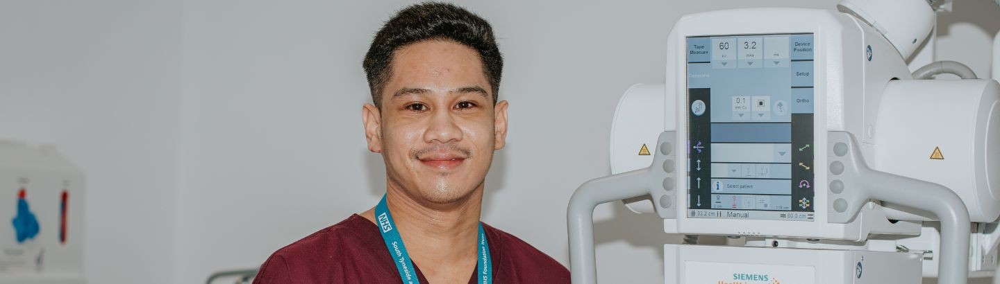
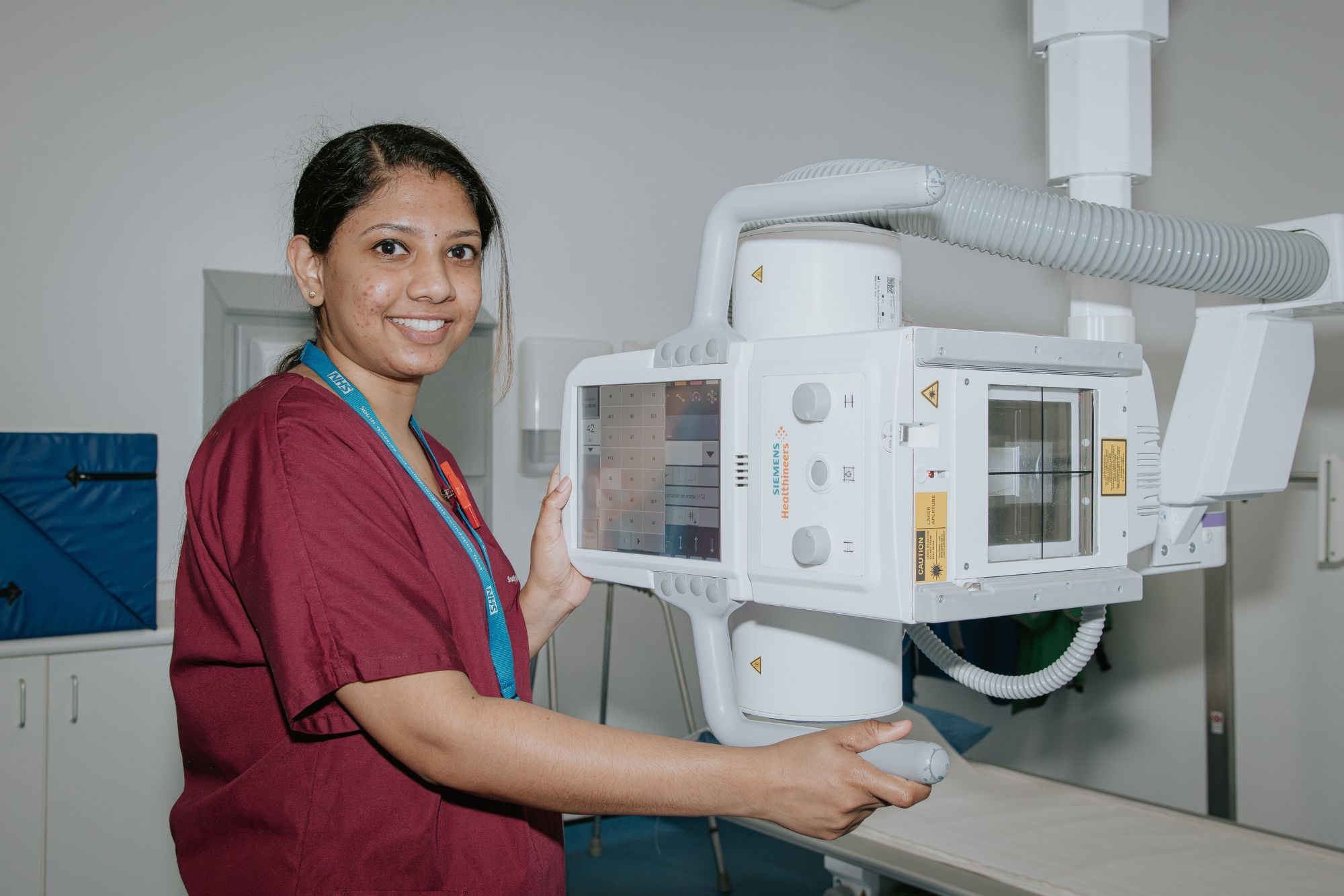
This is an x-ray:
A fluoroscopy is a type of X‑ray that shows moving pictures of the inside of your body in real time.
It is like watching an X‑ray video instead of just taking a single picture. Doctors use fluoroscopy to see how organs move, to guide tubes or needles, or to watch how a medicine or dye travels through the body.
The most common fluoroscopies are:
- Digital spot imaging - this is a type of X‑ray picture taken at one moment in time to show a clear, detailed view of a body part
- Digital subtraction angiography (DSA) - this is a special type of X‑ray that shows blood vessels. A dye is put into the blood, and the computer removes background images so the blood vessels are easier to see. This helps doctors check for blockages or other problems
What happens during a fluoroscopy test?
Many people feel nervous before the test, but it is safe, painless and we are here to support you.
Before the test
- A radiographer will greet you and explain what will happen
- You can ask questions at any time
- You may need to change into a gown or remove metal items like jewellery
- For some types of fluoroscopy, you may need to swallow a special drink or have a small amount of contrast given through an injection. This helps the pictures show up clearly
During the test
- You will lie on a table or stand, depending on the area being looked at
- A large camera will move above you. It may look big, but it will not touch you
- The radiographer may ask you to hold your breath for a few seconds or change position
- You may see the pictures on a screen if the team shows you
- The staff will talk to you throughout, so you always know what is happening
How it feels
- The test does not hurt
- You may feel warm, full or a bit strange if you swallow or receive contrast, but this feeling passes quickly
- Most fluoroscopy tests take about 10–20 minutes, but some may take a little longer
After the test
- You can usually go home or return to your ward right away
- You can eat and drink normally unless the staff tell you otherwise
- A doctor will look at the images and share the results with your care team
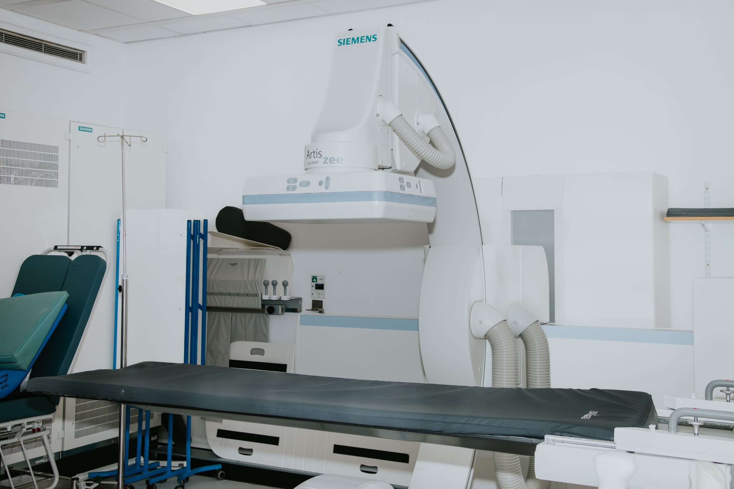
This is a fluoroscopy:
CT stands for computerised tomography.
A CT scanner uses X‑rays to take cross‑section pictures of the body. You can think of these pictures as thin slices of the area your doctor wants us to check.
CT scans are used to look at the chest, abdomen, pelvis, and brain. They can also help with orthopaedic problems. CT scanners can make clear pictures of the blood vessels, the heart and the bowel.
What happens during a CT scan?
Many people feel nervous before the scan, but the process is safe, quick and painless.
Before the scan
- A radiographer will meet you and explain what will happen
- You can ask questions at any time
- You may need to remove metal items like keys or jewellery
- Sometimes you may be given a special drink or injection to help the pictures show up better. We will tell you if this is needed
During the scan
- You will lie on a comfortable table that moves slowly into a short, round scanner
- The scanner is open at both ends, so you are not in a long tunnel
- You will be able to see the room around you
- The machine will make soft whirring sounds, but it will not touch you
- The radiographer will be in the next room, but they can see you the whole time and can talk to you through a speaker
- You can talk back if you need help
How it feels
- The scan does not hurt
- You just need to lie still for a few minutes
- Most scans take around 10 minutes or less
- If you have an injection, you might feel warm for a few seconds. This is normal and passes quickly
After the scan
- You can go home or return to your ward right away unless we tell you otherwise
- You can eat and drink normally
- A doctor will look at the pictures and share the results with your care team
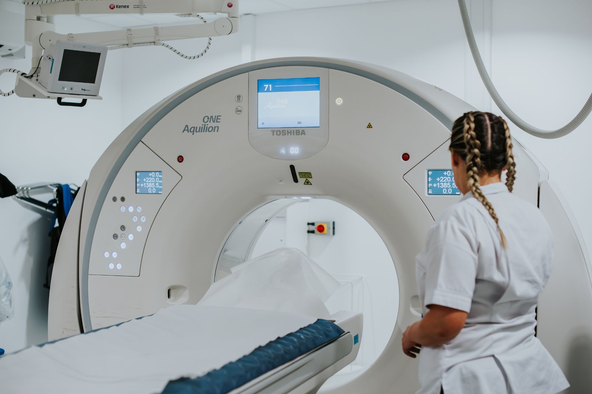
This is a CT scanner:
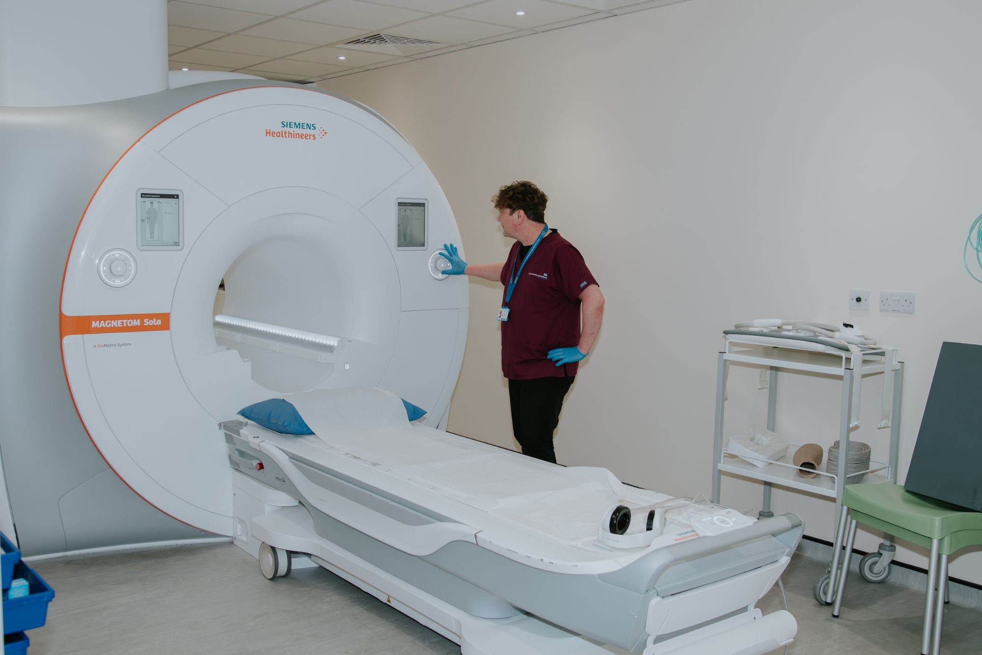
MRI stands for magnetic resonance imaging.
It uses a strong magnet and radio waves to take cross‑section pictures of the body without using X‑rays. It gives very detailed images of the soft parts inside the body.
While an ordinary X‑ray shows bones well, an MRI scan can show the brain, muscles, ligaments, nerves, blood vessels, and organs.
What happens during an MRI scan?
Many people feel nervous before the scan, but the test is safe, painless and we are here to help you feel comfortable.
Before the scan
- A radiographer will talk to you and explain what will happen
- You can ask any questions you have
- You may need to remove metal items like jewellery, keys, or belts
- You might be asked to change into a gown
- If you feel worried about small spaces, tell the staff. They can support you throughout the scan
During the scan
- You will lie on a padded table that slides into the scanner
- The scanner is a short tunnel-shaped machine
- The radiographer will make sure you are comfortable, and you will be given a button to press if you need help
- The radiographer will go to the next room, but they can see and hear you at all times, and they can talk to you through a speaker
- The machine makes loud tapping or buzzing sounds, but this is normal. You may be given earplugs or headphones
How it feels
- An MRI does not hurt
- You must lie still so the pictures are clear
- Some people find the space a bit tight, but you are never alone, and you can talk to the radiographer at any time
- Most scans take 15–30 minutes, depending on the area being looked at
After the scan
- You can go home or return to your ward straight away
- You can eat and drink normally
- A doctor will look at your images and share the results with your care team
This is an MRI scanner:
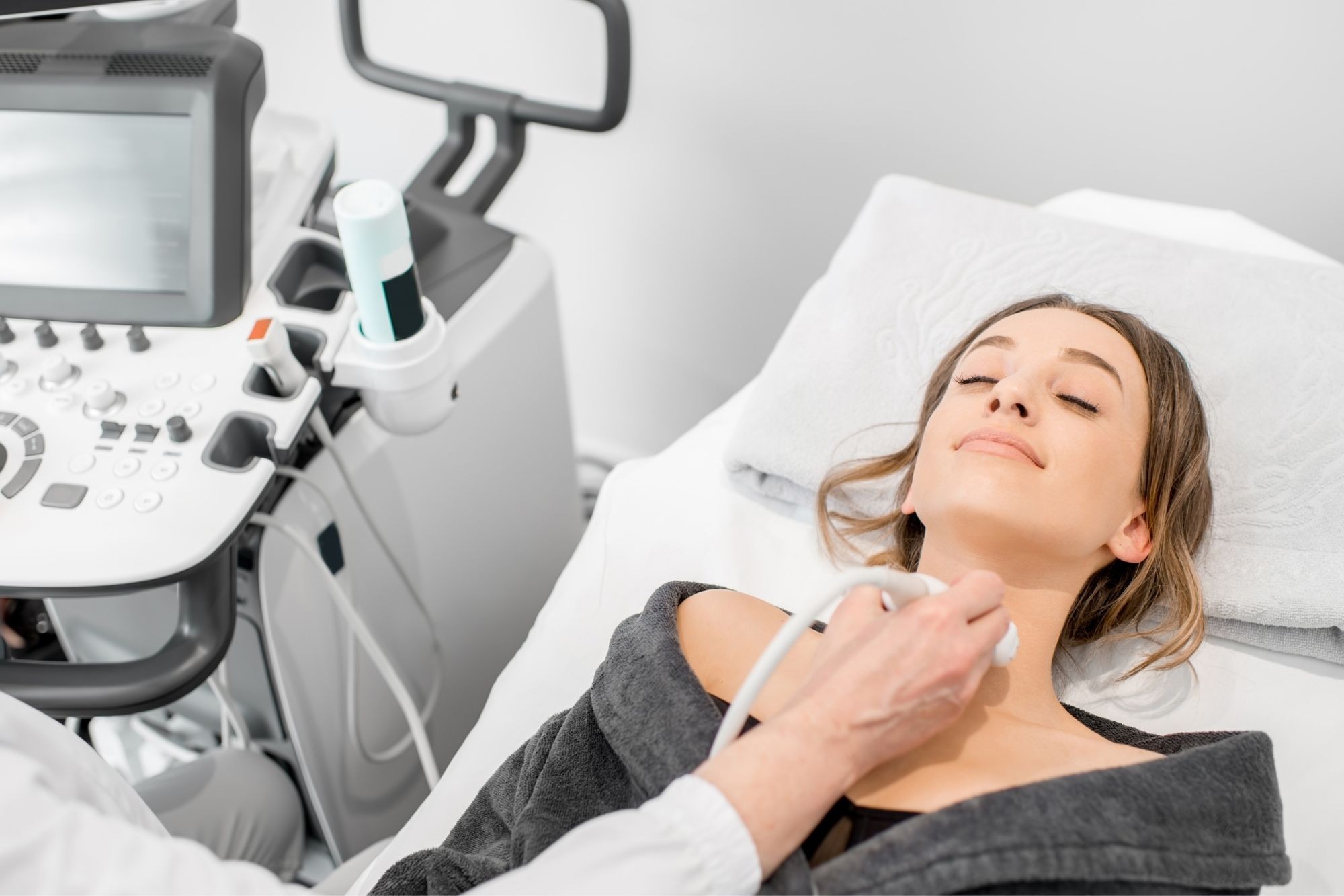
Ultrasound uses high‑frequency sound waves, not X‑rays, to make pictures of the inside of the body.
It is used to look at organs in the abdomen and pelvis, and it can also show soft tissues in the neck. Ultrasound is helpful for people with muscle or joint problems too.
A special type of ultrasound called Doppler is used to check for deep vein thrombosis and to look at how well blood flows through the arteries.
We do not do ultrasounds if you are pregnant in radiology. That will be done in maternity.
What happens during an ultrasound scan?
Many people feel nervous before the scan, but it is safe, gentle and does not hurt.
Before the scan
- A sonographer (the person who does the scan) will explain what will happen
- You can ask any questions at any time
- You may need to lift or remove clothing from the area being scanned, but you will be given privacy
During the scan
- You will lie on a bed in a comfortable position
- The sonographer will put a warm gel on your skin. This helps the sound waves make a clear picture
- They will move a small handheld device, called a probe, over the area being checked
- The probe does not hurt, and the pressure is usually gentle
- You may see the pictures on the screen if the sonographer shows you
- The room is usually quiet, and the scan takes about 10–20 minutes
How it feels
- Ultrasound does not cause pain
- The gel may feel a little cold at first
- You might feel slight pressure, but it should not be uncomfortable
- Staff will talk to you throughout the scan so you always know what is happening
After the scan
- The gel will be wiped off and you can get dressed
- You can go home or return to your ward right away
- You can eat and drink normally
- A doctor will review the images and share the results with your care team
This is an ultrasound:
Listen to our episode of Our People Podcast

Where can I find out more?
You might visit radiology in a number of different places.
South Tyneside
At South Tyneside District Hospital, we are based in the main building. Use the Ingham Wing entrance and follow signs to radiology.
We also do scans and tests at the Integrated Diagnostic Centre (IDC). This is the building in front of Ingham Wing at South Tyneside District Hospital.
Sunderland
Most of our main services are at X-ray in the Hylton Road block of Sunderland Royal Hospital.
We are on C Floor. Take the stairs or lifts from B Floor or through Chester Wing Outpatients Entrance 5.
Community sites
We also offer services at community sites including Galleries Health Centre in Washington and Palmer Community Hospital in Jarrow.
Our senior team in Radiology
Divisional director - Hannah Davidson
Directorate manager - Shauna Roberts
Clinical director - Ben Hall
Matron - Julie Mills