Throughout your cancer journey you may have some tests. This is to help us find out what is wrong with you. This will help us to find the best support and treatment for you.
Your tests will checked by a doctor. You should get your results within 1 or 2 weeks. You won't get any results at the time of the test. You can contact the hospital if you have not heard anything after a couple of weeks.
Waiting for results can make you anxious. You might have the contact details for a specialist nurse. You can contact them for information and support if you need to. It can help to talk to a close friend or relative about how you're feeling and the worries that you have.
Please click on the headings below for information relating to tests you may need to have.
What is an MRI scan?
An MRI scan uses magnetism to build up a picture of the inside of your body. MRI stands for magnetic resonance imaging.
Why might I need an MRI?
Doctors might use an MRI scan to show:
- If a lump or abnormal area is cancer or not.
- The size of the cancer and if it has spread.
Having your MRI
Before the scan the hospital will send you information. This is so you know what to expect.
- If you are having a scan of your tummy (abdomen), you might be asked to not eat anything for a few hours before the scan. This helps to get a clear picture.
- Some people feel a bit claustrophobic (a fear of confined spaces) in the scanner. If you are worried about the scan, you may be able to have a sedative. This will help you relax while you are being scanned. You should ask your GP or doctor about a sedative before you go for the scan.
The scan uses magnets so we need to be careful with metal. This is why the radiographer will go through a checklist.
This will talk about:
- About any metal implants you have. This could include surgical clips, bone pins, artificial joints and heart valves.
- About electrical devices. These can include a pacemaker, implanted defibrillators, nerve simulators or an cochlear implant.
- We want to know if you have ever worked with metal or in the metal industry. This is because very tiny fragments of metal can sometimes lodge in the body, especially the eyes.
- You should tell your radiographer if you are pregnant or think you could be.
- You can usually wear your own clothes if they do not have metal zips or buttons. Don't worry if you do not have clothes without metal, you can change into a gown.
Having metal in your body does not necessarily mean you cannot have an MRI scan. Your doctor and radiographer will decide if the MRI scan is safe for you. If you are not able to have an MRI scan, another type of scan can be used.
During the scan
You will be asked to lie very still on a bed. The bed moves slowly through the middle of a cylinder tube.
Some people are given an injection of a dye into a vein in the arm during the scan. It does not usually cause any discomfort. The dye is called a contrast medium. It helps certain types of tissues to show up more clearly on the scan. It is only given when necessary.
The radiographer leaves the room during the scan, but can see you through a screen. You will be able to talk to them through an intercom while you are having the scan.
Here are some points that tell you about the scan:
- The scan is not painful, but lying still on the bed during the scan can be a bit uncomfortable. It usually takes between 15 minutes and an hour.
- The scanner is very noisy and you will be given earplugs or headphones. It may be possible to listen to music during the scan.
- You will need to lie still on the bed as any movement can affect your results. If you get uncomfortable, let the radiographer know.
- Some people find it helpful to close their eyes while they are in the tunnel.
- Some tattoos contain metal, especially those with red dye in them. These can cause a warm or sometimes burning feeling during the scan. This is only in the area of skin where the tattoo is. If this happens, let the radiographer know straight away.
After the scan
Most people are ready to go straight home after their test.
If you have had a sedative:
- You will need someone to collect you from the hospital.
- You should have someone with you for 12 hours.
- You should not drive for 24 hours afterwards.
PLEASE NOTE: If you have a pacemaker or defibrillator you might still be able to have an MRI scan. The Radiology and Cardiology teams will need to check if it will be safe. Not all devices can be scanned.
If your device is safe, you will be booked for your MRI at the South Tyneside MRI Department. You cannot have this scan at the Integrated Diagnostic Centre or at Sunderland Royal Hospital.
What is a CT scan?
A CT scan makes a three-dimensional (3D) picture of the inside of the body. The 3D picture is built up using lots of detailed x-rays taken by the CT scanner.
It uses a small amount of radiation.
This is very unlikely to harm you.
It will not harm anyone you come into contact with.
Preparing for your scan
You will be given information about what you need to do before your scan. This will be things like:
- You may be asked not to eat or drink for a few hours before the scan. This depends on the part of the body being scanned. If this is a problem for you, call the number on your appointment letter.
- When you arrive at the hospital, you may be asked to put on a hospital gown.
- You may be asked to remove any jewellery or objects containing metal, including:
- Piercings
- Hair clips
- Zips
- Bra
You may be asked to have a drink or an injection of a dye. This is called contrast. It helps show certain areas of the body more clearly. The contrast may make you feel hot all over for a few minutes. It is important to let your doctor know if you are allergic to iodine or have asthma. This is because you could have a more serious reaction to the injection.
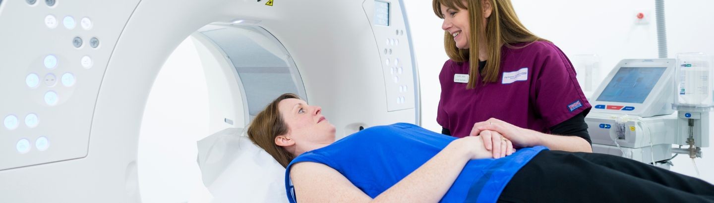
Having a CT scan
You have the scan in the x-ray department at the hospital. The person who works the scanner is called a radiographer. The scan takes 5 to 10 minutes, but you may be in the department for longer. You lie very still on a narrow bed.
The bed moves slowly back and forward through the doughnut-shaped machine. You will probably be able to go home as soon as the scan is over. The radiographer may suggest you drink plenty of water for the rest of the day. This will help flush out the dye (contrast).
What is a biopsy?
A biopsy is the removal of a small piece of tissue or a sample of cells. This is so they can be looked at under a microscope.
Biopsies are helpful to:
• tell if an abnormal area or lump is a cancer or not.
• to give your doctors information about the cell the cancer developed from.
• to help the doctors to plan the best treatment for you.
Biopsies can be done
• by the doctor in clinic
• in the x-ray department using ultrasound or CT scan to be used as a guide.
• in the operating theatre
• endoscopically (using an internal camera)
Sometimes it is not possible to do a biopsy. This could be because the area is too difficult to reach or you may be too unwell to have this done. Your doctor will discuss this with you in more detail. They may arrange for other tests to be done instead.
What happens during a biopsy?
How the biopsy is done will depend on where the sample is being removed from. Your doctor will explain this to you. You may be given a local anaesthetic to numb the area. A general anaesthetic may be given if a biopsy is taken in theatre.
After the biopsy you may have one or two stitches. You will have a dressing applied to the area to keep it clean.
Types of biopsy
There are different types of biopsy. Your specialist will explain which is best for you. The type of biopsy you have will depend on where in your body the abnormal area is. It may also depend on the type of cancer that is suspected.
The main types of biopsy are:
• fine needle aspiration (FNA)
• core biopsy
• open biopsy
Sometimes a small piece of tissue is removed (incisional). It could mean an entire lump or suspicious area (excisional) is taken away. The tissue is then examined under a microscope.
Bone Marrow Biopsy
Bone marrow is a spongy material inside your bones. Blood cells are made in your bone marrow.
A bone marrow biopsy is often needed if:
• Your doctor thinks you may have a problem with how your blood cells are made
• If you have a blood disorder, sometimes bone marrow biopsies are taken to check how well your treatment has worked.
What to expect
A bone marrow biopsy takes a small sample of the bone marrow. This is from within your hip bone.
You will be asked to visit the hospital for your biopsy. They are often done in the Phoenix Unit. They can also take place in the Outpatients department or on a ward, if you are already staying in hospital.
You will be helped to get comfortable on your side. Local anaesthetic will be used to numb the area.
A needle is put through your skin and into the hip bone. A small sample of liquid bone marrow is taken out. This is called a bone marrow aspitate.
They may also need to take a small sample of the core of bone marrow. This is called a trephine biopsy. This is taken in the same way. It uses a slightly larger needle.
It may feel a little uncomfortable for a few seconds when the liquid is taken from the bone marrow.
After a bone marrow biopsy
A small dressing will be put over the biopsy area. This can be taken off after 24 hours.
Most patients are ready to go home after their test.
Most patients do not need sedation.
If you do need sedation:
• You will need someone to collect you from hospital.
• You should have someone with you for 24 hours.
• You should not drive for 24 hours afterwards.
Click here to read more.
What is a sentinel lymph node biopsy?
A sentinel lymph node is the first lymph node to which cancer cells are most likely to spread from a primary cancer. Sometimes, there can be more than one sentinel lymph node.
A sentinel lymph node biopsy (SLNB) helps identify it. It removes it and looks at to see if cancer cells are present. It is used in people who have already been diagnosed with cancer.
A negative SLNB result shows cancer has not yet spread to nearby lymph nodes or other organs.
A positive SLNB result shows cancer may have spread to other nearby lymph nodes. This can help a doctor determine the stage of the cancer (extent of the disease within the body). It helps them develop a treatment plan.
What happens during an SLNB?
First, the sentinel lymph node (or nodes) must be found. A doctor injects a radioactive substance, a blue dye, or both near to the cancer. The surgeon uses a device to detect the lymph node that contains the radioactive substance or that is stained with the blue dye. The doctor makes a small cut in the skin. They remove the node.
The sentinel node is then checked for cancer cells. This is done by a pathologist. If cancer is found, the doctor may remove more lymph nodes. This could be during the same operation or at a different time. SLNB may be done on a day visit to hospital. It could mean a short stay in hospital.
SLNB is usually done at the same time the cancer is removed. But in some cases is done before that operation.
What are the benefits of SLNB?
SNLB helps doctors stage cancers. They help estimate the risk tumor cells have been able to spread to other parts of the body. If the sentinel node is negative for cancer, a patient may be able to avoid more lymph node surgery. This lessens the chance of complications lined to having many lymph nodes taken out.
What are the possible harms of SLNB?
All surgery to remove lymph nodes can have harmful side effects. Removal of fewer lymph nodes is usually associated with fewer side effects. The potential side effects include:
• Lymphedema, or tissue swelling. During lymph node surgery, lymph vessels leading to and from the sentinel node or group of nodes are cut. This changes the normal flow of lymph through the area. This may lead to an abnormal build up of lymph fluid and cause swelling. Lymphedema may cause pain or discomfort in the affected area. The overlying skin may become thickened or hard.
• The risk of lymphedema increases with the number of lymph nodes removed.
• Seroma, or a mass or lump caused by the build up of lymph fluid at the site of the surgery
• Numbness, tingling, swelling, bruising, or pain at the site of the surgery, and an increased risk of infection
• Difficulty moving the affected body part
• Skin or allergic reactions to the blue dye used in SNLB
• Rarely a false-negative biopsy result. This may give the patient and the doctor a false sense of security about the extent of cancer.
Is SLNB used to help stage all types of cancer?
No. SLNB is most commonly used to help stage breast cancer and melanoma. It is sometimes used to stage penile cancer and endometrial cancer. It is being studied with other cancer types.
Sentinel Node Biopsy for Cancer of the Penis
What is a mammogram?
A mammogram is an x-ray of your breasts. X-rays use high energy rays to take pictures of the inside of your body. Breast screening with a mammogram can help to find breast cancers early when they are too small to see or feel.
Having your mammogram
The health professionals who take mammograms are called mammographers. Often the mammographers are female. The mammogram only takes a few minutes, but the appointment may last about 30 minutes.
The mammographer positions one breast at a time between two flat plates on the machine. This helps to give a clear picture.
The mammographer takes the x-ray from behind a screen. This is to protect her from the radiation because she is taking x-rays everyday.
They take two pictures of the breast, one from above and one from the side. This repeated on the other breast.
You may have this test alongside other tests such as a breast examination and breast ultrasound. This happens in the same appointment. This is often called a triple assessment.
Some people find mammograms a bit painful. Most only feel mild discomfort. Either way, it doesn’t last for long.
Click here to read a leaflet with more information: Breast screening and the Assessment Unit
You can find leaflets in other languages about breast screening here: Your guide to NHS breast screening
What is a bone scan?
A bone scan is a special test used to look at the bones. It is a nuclear radiology test performed in the medical physics department.
- It is done to look for areas of change in bone.
- Or to see how well treatment is working.
Preparing for your bone scan
Before your scan you will have an injection of a small amount of radioactive substance.
- This helps to show up any abnormalities in the bone.
- This is not enough to be harmful.
- After the injection you can leave the department for a couple of hours. This allows the substance time to get to your bones.
- The nurse will tell you when to return to the department.
- While you are away drink a couple of extra pints of water to flush the injection through your body.
- Before the scan you will need to empty your bladder.
During your bone scan
You usually wear your own clothes for the scan. Sometimes you may be asked to change into a hospital gown.
You will be asked to empty your pockets and remove any metal objects. These can include keys, coins, belts, braces and jewellery.
You lie will down on a couch and have to keep very still while you go through the scanner.
The scan can take between 30 to 60 minutes, but you'll be at the hospital for several hours.
After your bone scan
The small amount of radioactivity left in your body disappears within the next 24 hours. In this time you should avoid close contact with babies, children and pregnant women.
The results of your bone scan may show changes called hot spots. These are not always cancer. Bone changes can happen for other reasons. These can include arthritis or previous broken bones. Your doctor will advise if any further tests are needed.
What is an endoscopy?
An endoscopy where organs inside your body are looked at using an instrument called an endoscope. This is is a long, thin, flexible tube. It has a light and camera at one end. Images of the inside of your body are shown on a television screen.
Endoscopy looks inside your body from an opening, This is most commonly the mouth. An endoscope can also be put inside the body through a small cut (incision) made in the skin. This is when keyhole surgery is being done. This test may have other names depending on the area examined.
Click here to watch a tour of our Endoscopy Unit
Different types of endoscopy
A bronchoscopy looks inside your windpipe (trachea) and bronchi (tubes going into lungs).
A laryngoscopy looks inside the part of the throat called the larynx.
A nasendoscopy looks through your nostril at the back of the mouth, nose and throat.
A hysteroscopy looks inside the womb. This is if there are problems such as irregular bleeding.
A flexible sigmoidoscopy looks inside the lower part of your large bowel.
A cystoscopy looks inside the bladder. This is if there are problems like urinary incontinence or blood in your pee.
An OGD (Oesophagogastroduodenoscopy) is often called a gastroscopy or gastrointestinal endoscopy. It looks inside the gullet (oesophagus), stomach and duodenum (the first part of the small bowel).
A enteroscopy looks further into the small bowel to the jejunum and ileum.
There are also Colonoscopy and Colposcopy which you can find information on the tests and scans page.
Why would I need to have an endoscopy?
An endoscopy can be used to:
- investigate unusual symptoms
- help perform certain types of surgery
- remove a small sample of tissue to be looked at more closely. This is called a biopsy.
After an endoscopy
Most people are ready to go home a couple of hours after their test.
If you have had a sedative:
- You will need someone to collect you from the hospital.
- You should have someone with you for 12 hours.
- You should not drive for 24 hours afterwards.
If you do not have a sedative, you can go home soon after you have had an endoscopy.
What is a colonoscopy?
A colonoscopy is a way of looking at the inside of your bowel. A doctor or nurse passes a thin flexible tube (colonoscope) into your back passage. The tube has a tiny light and camera on the end. This helps show up any abnormal areas.
A colonoscopy can also see whether there are any small growths (polyps) in the lining of the bowel. It is one of the main tests used to diagnose bowel cancer. This test is usually done in the hospital's endoscopy department. It takes about 30 minutes.
During the check, photographs and samples (biopsies) of the cells on the inside of the large bowel can be taken. If polyps are seen these can often be painlessly removed. This is done using a wire loop passed down the colonoscope.
Preparing for a colonoscopy
The bowel has to be completely empty for a colonoscopy. You will be given some information about what you need to do.
This will involve:
• Following a careful diet for a few days before your test.
• Taking laxative. The hospital will give you with instructions about when to take them.
Having your colonoscopy
Shortly before the colonoscopy, you may be given a sedative to help you feel relaxed.
Once you are lying comfortably on your side, the nurse or doctor will gently pass the colonoscope into your back passage. The tube is flexible. This is so it can easily pass around the curves of the bowel.
Having a colonoscopy can be uncomfortable. If you do have pain, it is usually mild. If you find it very painful, tell the doctor or nurse straight away. They may give you Entonox. This is a gas that can relieve pain. It is sometimes called gas and air. You breathe it in through a mouthpiece.
Sometimes it is not possible to see the whole bowel during a colonoscopy. This can happen if the bowel is not completely empty. Or it could be the colonoscope can't pass around a bend in the bowel to reach the end. If this happens, you may be asked to have another colonoscopy or a CT Colonography.
After a colonoscopy
Most people are ready to go home a couple hours after their test.
If you have had a sedative:
- You will need someone to collect you from the hospital.
- You should have someone with you for 24 hours.
- You should not drive for 24 hours afterwards.
If you only had Entonox you should be able to drive home when the nurses say you have recovered. You will not need someone to stay with you overnight.
It is rare to have any serious problems after the test. Contact your GP or go to your nearest emergency department (A&E) straight away if you:
- Have severe tummy pain.
- Have a high temperature.
- Being sick.
- Bleeding from the back passage.
Risks of a colonoscopy
Most people who have a colonoscopy have no problems, but rarely there can be problems.
A small number of people have heavy bleeding. This can be after having a polyp removed or a biopsy. This can usually be treated quickly during the test. However, some people may need to be admitted to hospital to have this treated.
Rarely, the bowel can be torn or damaged. If this happens, you will usually need an operation to repair the tear.
All the risks of a colonoscopy will talked about with your doctor. This will be part of the consent process.
Useful websites
Genetic and genomic testing
Genetic testing is sometimes called genomic testing. It finds changes in genes. These can cause health problems. It is mainly used to diagnose rare and inherited health conditions. It can also be used to diagnose some cancers.
To watch a short video regarding genetic testing click here.
Why would I be offered a genetic test?
You may be offered a test because:
• Your doctor thinks you might have a health condition caused by a change to 1 or more of your genes.
• Someone in your family has a health condition that's caused by changes to genes.
• Some of your close relatives have had a particular type of cancer. This could be inherited.
• You or your partner have a health condition that could be passed on to your children.
If you have any questions, talk to your doctor about having a genetic test.
What can a genetic test tell me?
Genetic tests are sometimes recommended to:
• Help to diagnose a rare health condition in a child.
• Help you understand whether an inherited health condition may affect you, your child or another family member. It can help you decide whether to have children.
• Show if you are at higher risk of getting certain health conditions. This can include some types of cancer.
• Guide doctors in deciding what medicine or treatment to give you.
• Guide doctors on whether you are able to join a clinical trial.
Who can have a genetic test on the NHS?
You need to be referred for genetic testing by a doctor. Talk to your hospital doctor about whether testing is right for you.
Genetic testing is free on the NHS if you are referred for it by a hospital doctor. You will only be referred if you have a suspected genetic health condition. Or it could be you have a particular type of cancer.
Genetic counselling
If you’re offered a genetic test, you may be referred to a genetic counsellor. This can help you think through what the test means for you and your family.
A genetic counsellor can help you understand:
• The risks and benefits of you having a genetic test
• The potential results of your test and what they mean
• How your family members may be affected if the test shows a serious health condition runs in your family
• The risk of you and your partner passing on a health condition to your children
• Your options if you have a child with an inherited health condition and you do not want your next child to inherit it
A genetic counsellor can also direct you to relevant patient support groups.
The impact on your family
You may want to think about how the results may affect you and your family. There is also a chance that the test gives you information about your relatives you or they may not have known.
For example, it may show that you were adopted. It could show your biological father is not who you thought they were. This is because the test can show you do not share genes with your family members.
Having a genetic test
A genetic test is usually done using a sample. This can be of your blood or saliva.
If you have been sent for a genetic test because you have cancer, the test will be on a sample of the tumour that has already been taken out as part of your treatment.
The sample is sent to a genetic laboratory to be tested.
Getting the results
You will be told when to expect the results of your test. Depending on the reason for your test, it could be weeks or months. You may need further tests.
The results from the test may show:
• You have a change in your genes which is known to cause a health condition
• You do not have a change in your genes which is known to cause a health condition
• It is not clear what the results mean for your health (but doctors may have a better understanding of the results in the future)
• after you get your results, you may be referred to a genetic counsellor to help you understand what they mean for you and your family.
How your data will be used
Your genetic data includes your sample of blood, tissue or saliva. It includes clinical information about your health condition. It also includes the results of your test.
This data may be used in:
• your care
• planning to improve the health services you and others receive
• research
Data from this testing is stored in a secure national database. When needed, information that can identify you is taken out. This can include your name and address. Your data can only be accessed by approved staff.
If you have a genetic test on the NHS, it is not possible to stop your data being stored and shared. If you have already opted out of data from your health records being shared, this does not apply to your genetic test data.
The NHS is responsible for your genetic data. Find out more about the NHS’s responsibility
Find out more about how the NHS manages your data in the NHS England Privacy Notice
Genetic testing in breast cancer
These links tell you about genetic testing in breast cancer. If you would like further information, please contact the Institute of Genetic Medicine at Centre for Life. It can be reached via Telephone 0191 2418600 or email geneticcounsellorenquiries@nhs.net
Breast Cancer Now -Genetic testing for altered breast cancer genes
Macmillan Cancer Support -Family history, genes and cancer risk
What is a PET-CT scan?
A PET stands for positron emission tomography. It is a special type of CT scan. It uses low-dose radioactive glucose (a type of sugar). This helps measure the activity of cells in different parts of the body. It shows where cells are more active than normal. This can indicate cancer.
PET scans are used to:
• Help find out if a tumour is cancerous (malignant) or non-cancerous (benign).
• Help find out if cancer has spread to other parts of the body.
• PET scans can also be used to examine any lumps that remain after treatment to see if they are scar tissue or whether cancer cells are still present.
Having your PET scan
A very small amount of a mildly radioactive substance is injected into a vein. This is usually in your arm. A scan is then taken a couple of hours later. There is a PET scanner at the Integrated Diagnostic Centre at South Tyneside District Hospital. You could also be asked to visit The Freeman Hospital in Newcastle or James Cook Hospital in Middlesborough for your PET scan.
This scan is only performed on patients undergoing Thyroid Cancer treatment.
It is undertaken post Radio iodine treatment. It helps to assess for additional thyroid cells in the body which where radio iodine uptake has occurred.